
Fig. 2
- ID
- ZDB-FIG-210203-10
- Publication
- Huang et al., 2020 - GFP expression pattern in pituitary and gonads under the control of nuclear progesterone receptor promoter in transgenic zebrafish
- Other Figures
- All Figure Page
- Back to All Figure Page
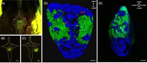
Cellular localization of green fluorescent protein (GFP) under the control of pgr promoter in the pituitary. A, The pituitary expressing GFP signals laid within a hypophyseal fossa. The white dash line indicated a cross‐shaped bone containing the pituitary. B, Dissected cross‐shaped bone (white dash line) containing the pituitary (red dash line). C, The intact pituitary (red dash line) was collected by breaking the bone (white dash line). D, Dorsal and, E, side view of the pituitary indicated the expression of GFP in the proximal pars distalis (PPD), and most of the GFP‐positive cells normally formed aggregates (red arrow), whereas a few were scattered as single cells (red arrowhead). The blue fluorescence represented the nucleus which was stained by Hoechst33342. Scale bars = 200 μm (A, B, C) and 50 μm (D, E). Pgr, progesterone receptor |