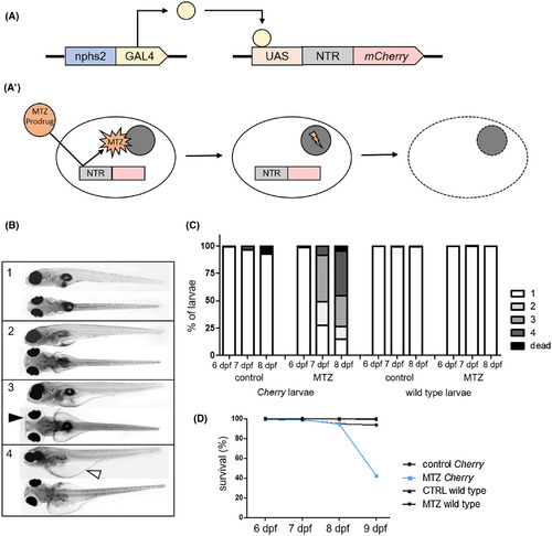
A, Schematically shows gene expression in the used transgenic zebrafish line. Gal4 is expressed under the nphs2 promotor. After binding to its specific recognition sequence UAS (upstream activation sequence), it activates the expression of a bacterial nitroreductase (NTR) and the red fluorescence protein mCherry. A′ shows a scheme of how MTZ is activated by NTR and subsequently induces DNA damage, which leads to apoptosis. B shows representative micrographs of 8 dpf zebrafish larvae to demonstrate the grading of developing edema after podocyte depletion. (1) no edema, (2) mild edema, (3) medium edema, and (4) severe edema. The black arrowhead exemplary indicates periocular edema, the blank arrowhead indicates abdominal edema. The graph in C shows relative distributions of phenotypes in n = 251 control‐treated and n = 275 MTZ‐treated Cherry larvae in four individual experiments and in n = 250 control‐treated and n = 250 MTZ‐treated wild‐type larvae. As visualized in the diagram in D, survival rates of podocyte‐depleted Cherry larvae are similar to controls until an abrupt decrease at 9 dpf
|
