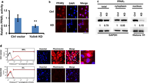
Effect of Yulink KD on PPARγ activity in HL-1 cardiomyocytes. a The DNA-binding activity of nuclear PPARγ in HL-1 cardiomyocytes was significantly reduced by Yulink KD using Yulink-shRNA (left panel); values normalized to those in cells treated with Ctrl vector (relative PPARγ activity) are shown. (n = 3, ** p < 0.01, Student’s t test). b The nuclear PPARγ expression level (Red) was decreased in Yulink KD cardiomyocytes. In the immunofluorescence assay, PPARγ was stained red, and nuclei were stained with DAPI (blue). c The amounts of total, cytoplasmic, and nuclear PPARγ were determined by Western blot, and normalized to an internal control (GAPDH or Histone H1) (right panel). Total PPARγ protein was decreased by 30%, while nuclear PPARγ was decreased by 85% in Yulink KD cardiomyocytes. d KD of Yulink decreases the uptake of the PPARγ ligand 15d-PGJ2-biotin in cardiomyocytes. Cells were incubated in 1 μM 15d-PGJ2-biotin for 3 h, and then fixed and stained with streptavidin-Alexa Fluor647 (red). Signals were detected using flow cytometry and immunofluorescence. Black lines indicate cells incubated without ligand; red lines indicate cells incubated with ligand in flow cytometric analyses. In the immunofluorescence assay, 15d-PGJ2-biotin was stained red, and nuclei were stained with Hoechst 33342 (blue). The majority of cardiomyocytes transfected with control vector took up the 15d-PGJ2-biotin (upper panel), while only 2% of Yulink KD cardiomyocytes took up the ligand (bottom panel)
|

