|
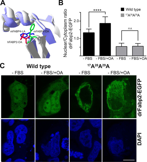
Wild-type and mutant drFabp2 ligand-dependent activation of nuclear traslocation.A) Superimposed structures of the α-helical regions of mFABP4 bound to linolenic acid (mFABP4-LA, PDB ID: 2Q9S), mFABP4 bound to oleic acid (mFABP4-OA, PDB ID: 1LID) and hFABP2 bound to oleic acid (hFABP2-OA, PDB ID: 2MO5) α-helical region. The side chain of the Phe residue implicated in ligand-dependent response to ligands is shown for FABP4 bound to linolenic acid (blue), mFABP4 bound to oleic acid (green) and hFABP2 bound to oleic acid (red). B) Graphical representation of nuclear/cytoplasmic fluorescence intensity relationship (mean ± SD) of wild- type and 17A28A29A drFabp2-EGFP in Caco-2 cells cultured in media without FBS (-FBS) or in media without FBS and with oleic acid (-FBS/+OA). Statistical significance is indicated, **** p < 0.0001, n.s. not significant. C) Representative single focal planes of Caco-2 cells transfected with wild- type or 17A28A29A drFabp2-EGFP (green) cultured in media without FBS (-FBS) or in media without FBS and with oleic acid (-FBS/+OA). Nuclei counterstaining with DAPI is shown in blue. Bar: 10 μm.
|