|
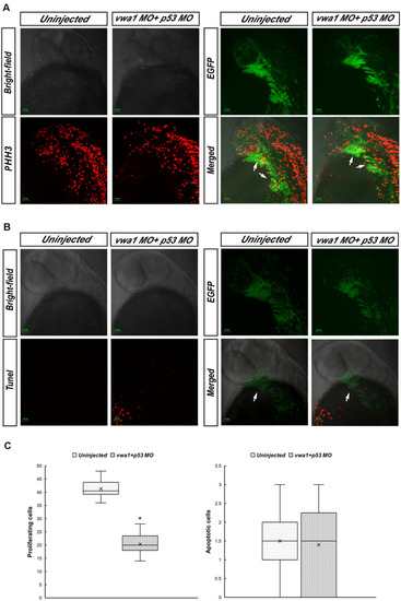
Proliferation and apoptosis of cranial neural crest cells at approximately 30 hpf. (A) Immunofluorescence results showing anti-phosphohistone H3 (PHH3) staining demonstrating cell proliferation. Merged image demonstrates higher anti-PHH3 signals in cranial neural crest cells compared with uninjected embryos. Arrows: pharyngeal arches. (B) Apoptosis assessed by immunofluorescence detection of TUNEL assay. The apoptosis signal in the vwa1 + p53 MO group did not coincide with EGFP fluorescence. Arrows: pharyngeal arches. (C) Comparison of apoptotic and proliferative numbers of neural crest cells in pharyngeal arches between vwa1 + p53 MO-injected embryos and uninjected control embryos. The difference in cell proliferation was statistically significant (p < 0.05, Student’s t-test). Lower/upper whiskers: 1st and 4th quartiles of the data; box: 2nd and 3rd quartiles of the data; mid-line: median of the data; ×: mean value of the data; *: significantly different.
|