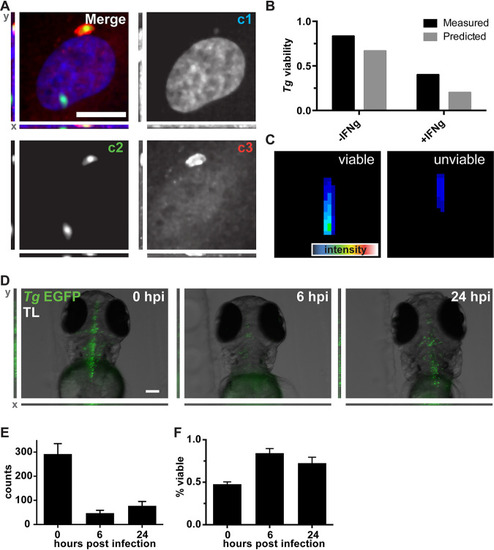
Mimicry embedding and weight transfer employed for Toxoplasma gondii (Tg) viability detection in cell culture and in vivo. (A) Merged three-channel fluorescent image of a HUVEC infected with T. gondii-EGFP. Individual channels represent DNA stain (c1), T. gondii-EGFP (c2), and ubiquitin (c3). A total of 2,694 images were obtained from 3 biological replicates. Bar, 25 μm. (B) Quantification of weakly labeled (measured) and CapsNet-inferred (predicted) viable and unviable parasites. (C) Representative reconstructions of the trained CapsNet network for viable and unviable classes of T. gondii-EGFP Z-profiles. (D) Representative images (maximum-intensity projections) of zebrafish (D. rerio) larvae infected with T. gondii-EGFP at 0, 6, and 24 hpi. The same 10 larvae were followed over time. Bar, 100 μm. (E) ZedMate-detected T. gondii counts at 0, 6, and 24 h postinfection. (F) In vivo inference of T. gondii-EGFP viability over time using the DropConnect viability model trained on in vitroT. gondii data. n = 10 Z-stack images per time point (3 biological replicates). Values are means and SEM. Statistical validation of machine learning models is provided in Fig. S5.
|
