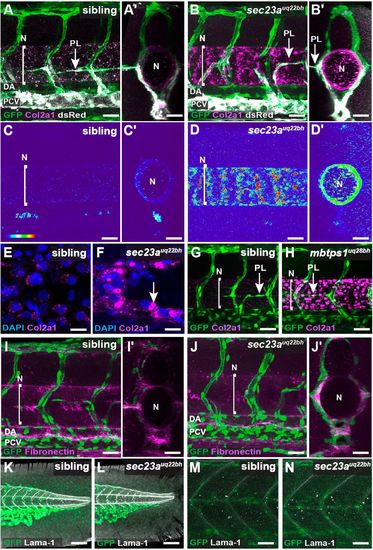
sec23auq22bh and mbtps1uq28bh mutant embryos display failed secretion of Collagen2a1. (A-D′) IF staining of Col2a1 and eGFP in 2 dpf Tg(fli1a:nEGFP); Tg(-5.2lyve1b:dsRed) siblings (A, cross-section A′) and sec23auq22bh mutants (B, cross-section B′) showing vasculature, secondary sprouts and Col2a1 labelled green, grey and magenta, respectively. Thermal maps showing lateral and cross sectional images reveal that sec23auq22bh embryos (n=10) exhibit an accumulation of Col2a1 around the notochord when compared with siblings (n=22) at 2 dpf (C-D′). (E,F) High magnification confocal images show intracellular accumulation of Col2a1 (indicated by white arrow) in NSCs in sec23auq22bh (F) when compared with siblings (E). (G,H) Confocal images as in A of siblings (G, n=12) and mbtps1uq28bh (H, n=6) mutants show similar accumulation of Col2a1 at 2 dpf as sec23auq22bh. (I-J′) Confocal images as in A showing vasculature and secondary sprouts (green) and Fibronectin (magenta) indicate no obvious defect in secretion of Fibronectin in siblings (I, cross-section I′; n=36) and sec23auq22bh embryos (J, cross-section J′; n=8). (K-N) Confocal images as in A showing vasculature (green) and Laminin (Lama1; grey) indicate no obvious defect in secretion of Laminin in the fin (K,L) or somite (M,N) when siblings (n=36) were compared with sec23auq22bh mutants (n=7). Line marked N indicates notochord. DA, dorsal aorta; N, notochord; PCV, posterior cardinal vein. Scale bars: 100 µm in A-F; 80 µm in G-N.
|
