
Fig. 2
- ID
- ZDB-FIG-200820-22
- Publication
- El-Nachef et al., 2020 - De novo enteric neurogenesis in post-embryonic zebrafish from Schwann cell precursors rather than resident cell types
- Other Figures
- All Figure Page
- Back to All Figure Page
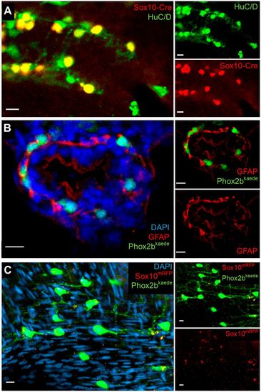
Further assays in larvae and adults support an absence of resident neuronal progenitors and enteric glia in the intestine. (A) Lineage tracing with an indelible Sox10-Cre line suggests enteric neurons are the sole fate of enteric vagal neural crest cells. At 5 dpf, fish were fixed and underwent IHC for the Cre reporter, mCherry and the neuronal marker HuC/D. All Cre-labelled cells colocalized with HuC/D, and no Cre+, HuC/D− cells were observed. (B) IHC with GFAP does not demonstrate convincing enteric glial cell bodies. Phox2b-kaede fish were fixed at 5 dpf, and axially sectioned for IHC for GFAP, a glial marker with cytosolic expression. Imaging of the endogenous Phox2b-kaede signal in concert with the GFAP IHC revealed a fibrillary pattern of GFAP closely associating with enteric neurons and other cells, which probably represents projections from extrinsic glia or nonspecific binding to other fibrillary proteins. (C) Whole-mount imaging of adult zebrafish intestine suggests that enteric glia and resident neuronal progenitors are not detectable by classical criteria later in development. Adult intestine from Phox2b-kaede×Sox10-mRFP fish that underwent optical clearing with RIMS revealed numerous enteric neurons, but no cell bodies expressing Sox10. Extrinsic glial projections are suggested by a fibrillary pattern of Sox10 expression. Scale bars: 10 µm. |