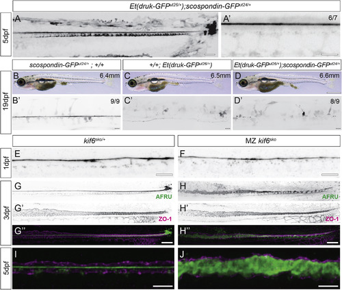
Loss of the Reissner Fiber Is Associated with Scoliosis in Additional Independent Scoliosis Mutant Zebrafish Strains (A and A’) Inverted grayscale maximal Z projections of confocal images of Et(druk-GFPdut26/+); scospondin-GFPut24/+ demonstrating typical assembly of the Reissner fiber, floor plate, and terminal ampulla expression in these mutant knockin embryos at 5 dpf. Note that GFP expression is contributed both by the Et(druk-GFPdut26/+) transgene insertion and from the scospondin-GFPut24/+ allele. Scale bars, 25 μm in (A) and 5 μm in (A’). (B–D) Bright-field images of a wild-type juvenile scospondin-GFPut24/+ (B), Et(druk-GFPdut26/+) (C), and Et(druk-GFPdut26/+); scospondin-GFPut24/+ (D) displaying the onset of mild scoliosis. Scale bar, 1 mm; standard length in upper right corner. (B’–D’) Inverted grayscale maximal Z projections of confocal images of the same fish in (B), (C), and (D) to highlight the spinal cord are shown. At 19 dpf, in wild-type scospondin-GFPut24/+, we observe high expression of the Reissner fiber (100%; n = 14 size range 6.3–6.8 mm; B’); in scoliosis mutant Et(druk-GFPdut26/+); +/+, the Reissner fiber is not labeled but some spinal cord cells express GFP (n = 6 size range 6.0–6.5 mm); and in double mutant Et(druk-GFPdut26/+); scospondin-GFPut24/+, we observed curvature of the spinal canal and a complete loss of a Reissner fiber (94%; n = 16 size range 6.1–7.0 mm). Scale bars, 25 μm. (E and F) Inverted grayscale maximal Z projections from confocal imaging of AFRU-stained zebrafish embryos at 1 dpf (30 hpf). The Reissner fiber is observed in heterozygous kinesin family member 6 (kif6sko/+) (E) and homozygous kif6sko mutants (F) (8/8; for each genotype). Scale bars, 10 μm. (G–H”) Inverted grayscale maximal Z projections of confocal imaging of AFRU- and ZO-1-stained zebrafish at 3 dpf. ZO-1 localizes to tight junctions and shows the location of the central canal epithelium. Heterozygous kif6sko/+ animals have an intact Reissner fiber in the central canal (13/13; G–G”), whereas in homozygous kif6sko, the Reissner fiber is disassembled and Reissner material fills the space of the central canal (12/13; H–H”). Scale bars, 25 μm. (I and J) Merge of maximal Z projections of confocal imaging of AFRU- and ZO-1-stained zebrafish at 5 dpf. Heterozygous kif6sko/+ animals have an intact Reissner fiber in the central canal (I), but in homozygous kif6sko animals, diffuse Reissner material fills the central canal lumen (J). Scale bars, 10 μm.
|
