
Fig. 6
- ID
- ZDB-FIG-200714-18
- Publication
- Sidhwani et al., 2020 - Cardiac function modulates endocardial cell dynamics to shape the cardiac outflow tract
- Other Figures
- All Figure Page
- Back to All Figure Page
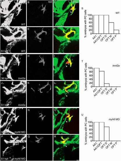
Cardiac function promotes addition of endothelial cells from the aortic arches to the OFT endocardium. (A-U) Three-dimensional reconstructions depict the photoconversion of a portion of AA1 endothelium at 36 hpf (as in Fig. 2Q-S) in wild-type (WT) (A-C), tnnt2a mutants (G-I) or myh6 morphants (M-O). By 51 hpf, labeled cells are routinely detected (as in Fig. 2T-V) in the WT OFT, as quantified in S (n=5; N=2). See also Movie 5. In contrast, in tnnt2a mutants (J-L) and myh6 morphants (P-R) at 51 hpf, labeled cells are found less often in the distal OFT (OFT D) and are entirely absent from the middle and proximal OFT (OFT M and OFT P, respectively), compared with WT (D-F), as quantified in T and U [n=6 (tnnt2a), 5 (myh6 MO); N=2]. See also Movies 6 and 7. Arrowheads indicate examples of cells that were not labeled by photoconversion (green only) and arrows indicate examples of cells that were labeled by photoconversion (red and green). (S-U) Bar graphs (as in Fig. S1) indicate the percentage of embryos in which labeling of AA1 endothelium at 36 hpf resulted in detection of photoconverted (PC) cells in AA1, at the junction of AA1 and the OFT (AA1/OFT), OFT D, OFT M or OFT P at 51 hpf. V, ventricle. Scale bar: 20 μm. |