
Fig. 4
- ID
- ZDB-FIG-200714-16
- Publication
- Sidhwani et al., 2020 - Cardiac function modulates endocardial cell dynamics to shape the cardiac outflow tract
- Other Figures
- All Figure Page
- Back to All Figure Page
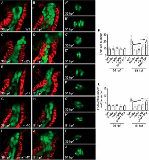
Cardiac function promotes accrual of endocardial cells in the OFT. (A-L) Lateral slices (A-J) and middle cross-sections (A′-J′) through the OFT (as in Fig. 1C,D) illustrate the number of OFT endocardial cells at 36 (A,C,E,G,I) and 51 hpf (B,D,F,H,J). As shown in Fig. 1S,T, the number of endocardial cells in the wild-type (WT) OFT increases between 36 (A,A′) and 51 hpf (B,B′), as quantified in (K,L). In contrast, in tnnt2a mutant embryos, OFT endocardial cells fail to accrue between 36 (C,C′) and 51 hpf (D,D′). A similar deficiency in OFT endocardial accrual is found in myh7 mutant embryos (E,E′,F,F′), and myh6 mutant embryos (G,G′,H,H′) also exhibit a reduction in OFT endocardial cell number by 51 hpf. However, gata1 morphants (I,I′,J,J′) exhibit a normal number of OFT endocardial cells. (K,L) Bar graphs indicate mean±s.e.m. [n=15 (WT), 8 (tnnt2a), 6 (myh7), 6 (myh6), 7 (gata1 MO) for 36 hpf; 24 (WT), 6 (tnnt2a), 10 (myh7), 13 (myh6), 8 (gata1 MO) for 51 hpf; N=2; ***P<0.001, ****P<0.0001; differences from WT at same stage, Student's t-test]. Scale bar: 5 μm. |