
Figure 4
- ID
- ZDB-FIG-200529-4
- Publication
- Parakh et al., 2020 - The Redox Activity of Protein Disulfide Isomerase Inhibits ALS Phenotypes in Cellular and Zebrafish Models
- Other Figures
- All Figure Page
- Back to All Figure Page
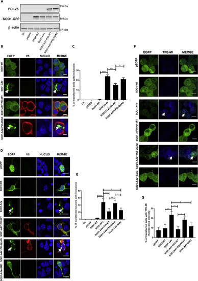
The Oxidoreductase Activity of PDI Is Protective against Inclusion Formation and Protein Unfolding in Mutant SOD1 expressing cells Immunoblotting was performed to confirm that similar transfection efficiencies were present and that co-expression of PDI-WT or PDI-QUAD did not alter the expression of SOD1. An anti-GFP antibody was used to detect SOD1-WT or mutant SOD1A4V (SOD1-A4V), in cells co-expressing empty vector pcDNA3.1 or PDI-V5 (WT or QUAD). β-actin was used as a loading control (bottom panel). (B) Immunofluorescence detection of EGFP in cells expressing SOD1-WT (row 1) or SOD1-A4V (inclusions represented by white arrows, row 2), co-expressed with PDI-WT or PDI-QUAD (rows 3, 4). (C) Significantly fewer cells formed inclusions when PDI-WT was co-expressed with SOD1-A4V (∗∗∗p < 0.001), and significant difference was observed between PDI-WT and PDI-QUAD expressing cells (∗p < 0.05). (D) Immunofluorescence detection of EGFP-positive inclusions present in mouse primary neurons co-expressing EGFP only (row 1), SOD1-WT (row 2) or SOD1-A4V (row 3), with PDI-WT or PDI-QUAD, or treated with BMC (rows 4, 5, 6). (E) Significantly fewer neurons formed inclusions when PDI-WT was co-expressed with SOD1-A4V (∗∗p < 0.01) or treated with BMC (∗p < 0.05). A significant difference was observed between SOD1-WT and mutant SOD1-A4V (∗∗∗∗p<0.0001) cells. n = 35, ANOVA followed by Tukey's post hoc test. (F) TPE-MI fluorescence in Neuro-2a cells expressing pEGFP (row 1), SOD1-WT (row 2), or SOD1-A4V cells (row 3), co-expressing PDI-WT or PDI-QUAD, or treated with BMC (rows 4, 5, 6). (G) Significantly fewer cells displayed TPE-MI fluorescence when PDI-WT was co-expressed with SOD1-A4V or treated with BMC (∗∗p < 0.01 and ∗p < 0.05). Scale bars: 10 μm in (B), 10 μm in (D), 15 μm in (F). |