FIGURE

Fig 1

ID
ZDB-FIG-200429-7
Publication
Arribat et al., 2020 - Spastin mutations impair coordination between lipid droplet dispersion and reticulum
Other Figures
All Figure Page
Back to All Figure Page
Fig 1

Spastin splice variants in zebrafish.

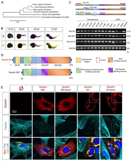
(A) Spastin phylogenic tree (designed from www.phylogeny.fr –Substitution model: Dayhoff). (B) Whole-mount in situ hybridization of Spastin transcript in zebrafish embryos (at 14, 18, 28, 72 hpf). Antisense probe (AS) correspond to Spastin transcript. Sense probe (S) to negative control. n = 10 embryos for each group. (C) RT-PCR amplifying different regions of Spastin from embryonic and adult zebrafish cDNA. (D) Schematic representation of Spastin splice variants in zebrafish. (E) Confocal microscopy images of zebrafish embryonic cells overexpressing Spastin splice variants (treated with 300μM oleic acid for 18h before acquisition). Top right insets correspond to higher magnification centered on LDs. Cherry-tagged Spastin appears in red. Tubulin labeling corresponds to microtubules (cyan), Bodipy to LDs (yellow) and Hoechst to nucleus (blue). N = 4 experiments. See also S1 Fig.

Expression Data
Gene:
Fish:
Anatomical Terms:
Stage Range: 1k-cell to Adult

Expression Detail
Antibody Labeling
Phenotype Data

Phenotype Detail
Acknowledgments
This image is the copyrighted work of the attributed author or publisher, and ZFIN has permission only to display this image to its users. Additional permissions should be obtained from the applicable author or publisher of the image. Full text @ PLoS Genet.