
Figure 2
- ID
- ZDB-FIG-200423-82
- Publication
- Murcia-Belmonte et al., 2019 - A Retino-retinal Projection Guided by Unc5c Emerged in Species with Retinal Waves
- Other Figures
- All Figure Page
- Back to All Figure Page
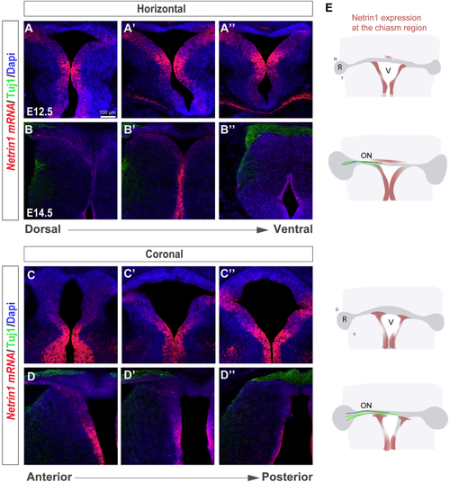
Netrin1 Is Expressed at the Developing Ventral Chiasm (A–D) Horizontal (A–B’’) and coronal (C–D’’) serial sections of E12.5 (A–A’’ and C–C’’) and E14.5 (B–B’’ and D–D’’) embryos at the level of the optic chiasm region stained by (E) Diagram summarizing the spatiotemporal expression of Netrin1 at the optic chiasm (red). RGC axons projecting to the brain (light green) or to the opposite optic nerve (dark green) are also represented. At E12.5, when RGC axons have not yet arrived at the chiasm, |