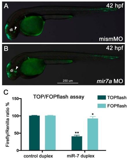
miR-7 negatively regulates Wnt signaling in zebrafish embryos and HEK293T cells. (A,B) miR-7 knockdown in zebrafish (mir7a MO) resulted in an increase of Wnt reporter expression in the ventral brain (B, green signals indicated by the white arrowhead) when compared with embryos injected with the control mismatch morpholino (mismMO) (A). Both panels display lateral views of 42 hpf Wnt:GFP transgenic embryos, anterior to the left; di: diencephalon. (C) HEK293T cells were co-transfected with TOPflash vector, NCDP, or 7DP, as well as Renilla luciferase control vector (pRL-TK). Similarly, the FOPflash vector (with mutated Tcf binding sites) was co-transfected with NCDP and 7DP, as well as pRL-TK. Luciferase activity was measured at 48 h post-transfection. About a 60% reduction in luciferase activity was observed in the TOPflash vector, whereas not much significant change was observed in the FOPflash vector. Luciferase readings were normalized to the co-transfected control Renilla luciferase vector pRL-TK plasmid. The error bars represent luciferase activity average ± SEM, where the experiment was done four times with two biological replicates each time (n = 6). Two-tailed t-tests are described as (**) where p < 0.01, and (*) where p < 0.05.
|
