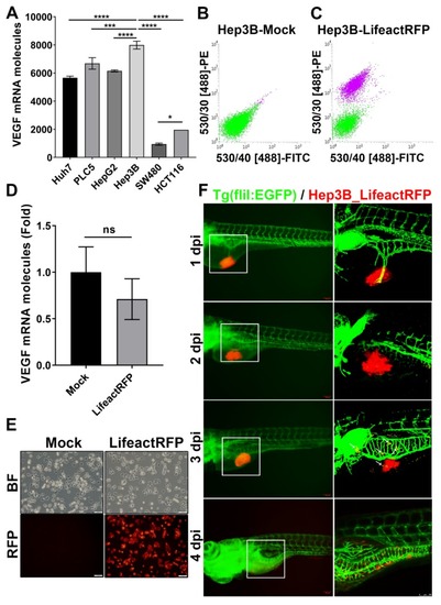
Establishment of stable RFP-expressing cancer cell line for tumor xenotransplantation study. (A) VEGF (VEGFA) mRNA expression in human hepatocellular carcinoma (Huh7, PLC5, HepG2, Hep3B) and colorectal cancer (SW480, HCT116) cell lines. The statistical significance was analyzed by one-way ANOVA. ns: non-significant, p > 0.05; *: 0.01 < p ≤ 0.05; ***: p ≤ 0.001; ****: p ≤ 0.0001; (B,C) FASC of un-transfected (B) and rLVUbi-Lifeact-TagRFP transfected Hep3B cells (C). The purple cell pools were sorted to create a stable Hep3B_Lifeact-RFP cell line. (D) mRNA expression of VEGFA in Hep3B and Hep3B_Lifeact-RFP stable line. ns, p > 0.05; unpaired Student’s t-test. (E) Representative bright field and fluorescent images of Hep3B and Hep3B_Lifeact-RFP stable line. Scale bar = 50 μm. (F) Representative images of Hep3B_Lifeact-RFP injected embryos from 1~4 day-post-injection (dpi) taken by fluorescent microscope (left) and confocal microscope (right). White arrow indicates metastatic cells at 3 dpi. Scale bar = 100 μm.
|
