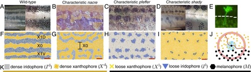
Self-organization during development. Diverse skin patterns form on zebrafish due to the interactions of pigment cells. (A) Wild-type zebrafish feature dark stripes and light interstripes (4, 11), while mutant patterns that form because a particular cell type is missing have altered, more variable patterns. (B) The nacre mutant (encoding mitfa) (9, 12) has an enlarged central orange region flanked by blue patches. (C) Pfeffer (encoding csf1rA) (9, 10, 13) is characterized by messy spots arranged horizontally (11). (D) Shady (encoding ltk) (11, 14) often features smooth black spots roughly arranged in stripes. Reproduced from ref. 11, which is licensed under CC BY 3.0. (E) Pigment cells extend long legs (measuring up to half a stripe width in distance) toward interstripe cells for communication (26). Reproduced from ref. 26, which is licensed under CC BY 3.0. (F–I) The agent-based model (20) replicates zebrafish patterns in silico. (Red scale bar, 500 μm throughout this paper.) The central light interstripe is labeled X0, and the next two interstripes are called X1V and X1D (11). (J) Rules for agent behavior in the model (20) depend on the cells in short-range disks and a long-range annulus. Reproduced from ref. 20, which is licensed under CC BY 4.0. (K) Summary of the main pigment cells involved in patterning. Interstripes consist of orange dense xanthophores and silver dense iridophores, and stripes contain yellow loose xanthophores, blue loose iridophores, and black melanophores.
|
