|
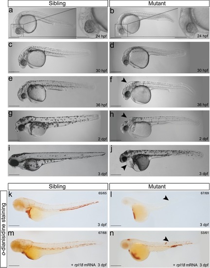
Rpl18-deficient embryos displayed physical abnormalities and anemia.a–j Phenotypes of wild-type siblings and rpl18−/− mutants (N = 7). At 24 hpf (a, b), aplasia of head region (dashed box) can be seen in mutant. At 30 hpf (c, d), pigmentation was delayed. At 36 hpf (e, f) and 2 dpf (g, h), a smaller head and edema (arrowhead) were present. At 3 dpf (i, j), edema (arrowhead) in heart and shorted tail extension was more apparent. Almost all embryos died at 4 dpf. k, lo-dianisidine staining showed depletion of erythroid cells in rpl18−/− embryos compared with wild-type siblings (arrowheads pointed to the sites of weak o-dianisidine staining, N = 5). m, nrpl18 mRNA (100 pg/embryo) injected to embryos, staining of 3 dpf mutants with o-dianisidine showed a partial recovery (N = 3). All scale bars represent 250 μm.
|