|
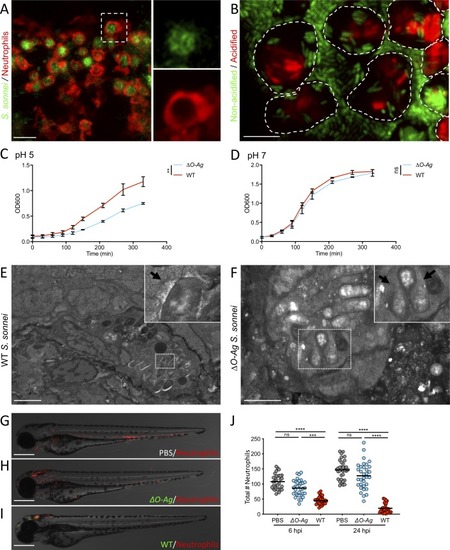
<italic>S</italic>. <italic>sonnei</italic> can resist phagolysosome acidification and promote neutrophil cell death in an O-antigen-dependent manner.A. S. sonnei is collected by neutrophils in large phagosomes. Larvae of the Tg(lyz:dsRed)nz50 strain (labelling neutrophils) were injected in the HBV with WT GFP-S. sonnei. Image taken at 3 hpi. Scale bar = 20 μm. B. S. sonnei is acidified in immune cell phagosomes. Larvae were injected in the HBV with WT GFP-S. sonnei which was also stained with pHrodo, a pH-sensitive dye that turns red in acidic environments. Image taken at 4 hpi, where GFP signal is attenuated for bacteria residing in acidified phagosomes (i.e. GFP is unstable and quenched at pH<6). Dashed lines highlight the outline of individual phagocytes. Scale bar = 10 μm. C,D. S. sonnei O-antigen contributes to acid tolerance in vitro. Growth curves of ΔO-Ag (blue) or WT (red) S. sonnei, cultured in tryptic soy broth adjusted to pH = 5 (C) or 7 (D). Statistics: unpaired t-test at the latest timepoint; ns, non-significant; **p<0.0021. E,F. S. sonnei requires the O-antigen to survive in phagosomes. Transmission electron micrographs of infected phagocytes from zebrafish larvae at 3 hpi with WT (E) or with ΔO-Ag (F) S. sonnei. E shows an intact phagocyte and S. sonnei residing within a phagosome (arrow points at phagosomal membrane). F shows that ΔO-Ag S. sonnei bacteria being degraded by a phagocyte (arrows point at region of major loss of bacterial cell integrity). Scale bars = 3 μm (E); 2 μm (F). G-J. The O-antigen is required for S. sonnei-mediated killing of neutrophils. Representative micrographs of larvae of the Tg(lyz:dsRed)nz50 strain injected in the HBV with PBS (G), GFP-ΔO-Ag (H) or WT (I) S. sonnei at 6 hpi and quantification of total neutrophil number at 6 and 24 hpi (J). Statistics: Kruskal-Wallis test with Dunn’s correction; ns, non-significant; ****p<0.0001. Scale bars = 250 μm.
|