|
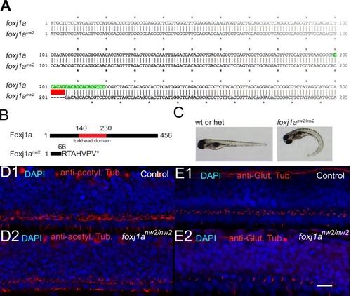
Characterization of cilia in the central canal of <italic>foxj1a<sup>nw</sup></italic><sup>2/<italic>nw</italic>2</sup> mutant embryo.(A) Alignment of the first 300 base pairs (bp) of the coding sequence of WT and mutant Foxj1anw2 allelle reveals that the Foxj1anw2 mutant allele carries a deletion of 5 bp from base pair 201 to 205. The deletion is indicated in red. The gRNA sequence is highlighted in green. (B) The Foxj1a protein consists of 458 amino acids and includes a DNA-binding forkhead domain (from amino acid 140 to 230, indicated in red). The Foxj1anw2 mutant allele results in a truncated protein due to a frame shift and early stop codon and lacks the forkhead domain. (C) - Foxj1anw2/nw2 homozygous mutant larvae display a curved body axis as seen on images of a control sibling (heterozygous or WT) versus a homozygous mutant larva taken with a stereomicroscope at four dpf. (D) Z-Projection stack of a lateral optical sections (Z-step = 1 µm over three sections) of the spinal cord immunostained with DAPI and against acetylated tubulin in a 30 hpf control sibling embryo (D1) and a foxj1anw2/nw2 curled-down embryo (D2). (E) Z-Projection stack of lateral optical sections (Z-step = 1 µm over three sections) of the spinal cord stained with DAPI and immunostaining against glutamylated tubulin in a 30 hpf control sibling (E1) and a foxj1anw2/nw2 curled-down embryo (E2). Scale bars are 15 µm.
|

