
Fig. 1
- ID
- ZDB-FIG-200124-52
- Publication
- Toms et al., 2019 - Missense variants in the conserved transmembrane M2 protein domain of KCNJ13 associated with retinovascular changes in humans and zebrafish
- Other Figures
- All Figure Page
- Back to All Figure Page
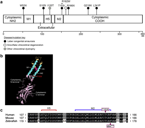
Location of reported (a) Schematic of the linear structure of Kir7.1 shows two transmembrane α helices (M1 and M2) with cytoplasmic NH2 and COOH termini, separated by an extracellular pore-forming loop that acts as a selectivity filter (H5). The location of published mutations is indicated and color-coded according to their associated disease. The missense mutation (p.Thr153Ile [T153I]) identified in families A and B in this study is highlighted with *. (b) Human Kir7.1 monomer model generated using Phyre2; the crystal structure of Kir3.2 was used as a template. The T153I mutation is highlighted in red. (c) Alignment of the human, mouse and zebrafish Kir7.1 protein sequences demonstrates the close proximity of the |
Reprinted from Experimental Eye Research, 189, Toms, M., Dubis, A.M., Lim, W.S., Webster, A.R., Gorin, M.B., Moosajee, M., Missense variants in the conserved transmembrane M2 protein domain of KCNJ13 associated with retinovascular changes in humans and zebrafish, 107852, Copyright (2019) with permission from Elsevier. Full text @ Exp. Eye. Res.