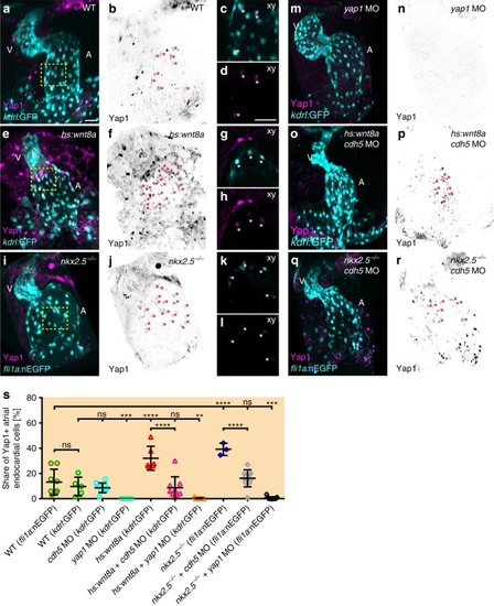
Yap1 nuclear localization within the atrial endocardium increases upon chamber expansion. a, e, i, m, o, q Reconstructions of confocal z-stacks of zebrafish hearts at 52 hpf expressing the endocardial reporters Tg(kdrl:EGFP)s843 or Tg(fli1a:nEGFP)y7 (cyan) and immunolabeling against zebrafish Yap1 (magenta). b, f, j, n, p, r Yap1 immunolabeling is inverted in black/white and endocardial cells which show co-localization with Yap1 labeling are marked with red asterisks. A atrium, V ventricle. Scale bars, 30 μm. c, d, g, h, k, l Magnifications of single confocal XY section planes (yellow box in a, e, i) are shown in c, g, k and, in d, h, l, only Yap1 immunolabeling is shown. Endocardial cells, labeled by Tg(kdrl:EGFP)s843 or Tg(fli1a:nEGFP)y7, that show co-localization with Yap1 are indicated with an asterisk. Scale bars, 30 μm. s Quantifications of the share of Yap1-positive endocardial cells relative to the total number of endocardial cells within the atrium. Upon Wnt8a overexpression (n = 6 hearts) or loss of Nkx2.5 (n = 3 hearts), the share of Yap1-positive endocardial cell numbers significantly increases within the developing atrial endocardium. Loss of Cadherin-5 (Cdh5) changes the share of Yap1-positive endocardial cells among Wnt8a overexpressing (n = 9 hearts) or nkx2.5vu179 mutant embryos (n = 10 hearts) to WT levels. Knock down of Yap1 in all conditions leads to a massive reduction of Yap1-positive endocardial cells within the atrium (yap1 MO: n = 11 hearts; yap1 MO + hs:Wnt8a: n = 9 hearts; yap1 MO + nkx2.5vu179 mutant: n = 11 hearts). Mean values ± SD are shown. One-way ANOVA was used to compare each condition with its WT control (ns not significant, **p < 0.01;***p < 0.001; ****p < 0.0001)
|
