
FIGURE
Figure 6
- ID
- ZDB-FIG-191230-798
- Publication
- Castro-Sánchez et al., 2019 - Functional analysis of new human Bardet-Biedl syndrome loci specific variants in the zebrafish model
- Other Figures
- All Figure Page
- Back to All Figure Page
Figure 6
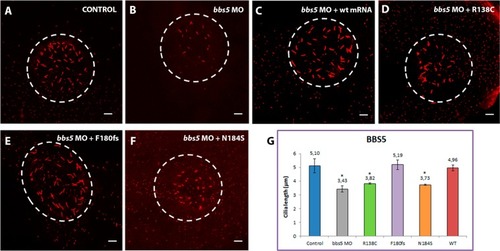
Representative images of KVs and comparison of average cilia length corresponding to |
Expression Data
Expression Detail
Antibody Labeling
Phenotype Data
| Fish: | |
|---|---|
| Knockdown Reagent: | |
| Observed In: | |
| Stage Range: | 5-9 somites to 10-13 somites |
Phenotype Detail
Acknowledgments
This image is the copyrighted work of the attributed author or publisher, and
ZFIN has permission only to display this image to its users.
Additional permissions should be obtained from the applicable author or publisher of the image.
Full text @ Sci. Rep.