|
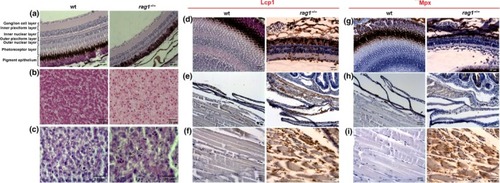
Rag1−/− zebrafish present with histological defects and alterations compared with wt siblings. (a–c) Premature aging signals in rag1−/− zebrafish. Representative images of retina (a) and liver (b, c) sections from 1‐year‐old wt and rag1−/− zebrafish (n = 5). (a) H&E‐stained eye sections revealed the loss of the photoreceptor layer and reduced thickness of nuclear layers. (b) The liver presents with hepatocyte cytoplasmic vacuolization. (c) Liver sections reveal numerous PAS‐positive areas, suggesting cytosolic accumulation of aging biomarker lipofuscin in hepatocytes (arrows). (d–i) Macrophage infiltration in rag1−/− zebrafish. Representative images of retina (d, g), skin (e, h), and muscle (f, i) sections from 1‐year‐old rag1−/− zebrafish. Consecutive anti‐Lcp‐ and anti‐Mpx‐immunostained sections reveal strong macrophage infiltration (Lcp+/Mpx‐) in the different tissues examined
|