FIGURE

Figure 2

ID
ZDB-FIG-191230-1883
Publication
Klingseisen et al., 2019 - Oligodendrocyte Neurofascin Independently Regulates Both Myelin Targeting and Sheath Growth in the CNS
Other Figures
All Figure Page
Back to All Figure Page
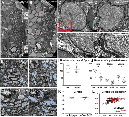
Figure 2

Ultrastructural Analyses of the nfascbue56 Mutant Spinal Cord

(A and B) Transmission electron microscopy (TEM) images of sections through the spinal cord of wildtype (A) and nfascbue56 mutants (B) at 5 dpf. Dorsal to the top, midline left. Asterisks show myelinated cell bodies. Scale bars, 5 μm.

(C–D′) TEM images of cell bodies in wildtype (C and C′) and ue56 mutants (D and D′) showing enwrapment of a cell body in the mutant with multi-lammellar myelin. Scale bars, 1 μm.

(E–H) High magnification views of myelinated axons in the dorsal (E and F) and ventral (G and H) spinal cord of wildtype (E) and mutant (F) animals. Unmyelinated axons with a diameter >0.3 μm highlighted in blue. Scale bars, 0.5 μm.

(I) Number of axons with a diameter > 0.3 μm in wildtype and nfascbue56 mutants (wildtype mean 114.5 ± 23.71 SD, n = 6 animals, nfascbue56 mutant mean 111.4 ± 23.69 SD, n = 7 animals, p = 0.8201, t test).

(J) Total number of myelinated axons in wildtype and nfascbue56 mutants, and numbers in the dorsal and ventral domains of the spinal cord. (Total: wildtype mean 92.17 ± 19.57 SD and nfascbue56 mutant mean 53.57 ± 18.20 SD, p = 0.0036, t test. Dorsal: wildtype mean 36.17 ± 7.89 SD and nfascbue56 mutant mean 21.93 ± 8.60 SD, p = 0.0103, t test. Ventral: wildtype mean 56.00 ± 12.52 SD and nfascbue56 mutant mean 31.64 ± 11.81 SD, p = 0.0041, t test. All sets wildtype n = 6 animals and nfascbue56 mutant n = 7 animals).

(K) Average g-ratio per animal in wildtype and nfascbue56 mutants at 5 dpf (wildtype mean 0.72 ± 0.01 SEM, n = 6 animals and nfascbue56 mutant mean 0.73 ± 0.01 SEM, n = 7 animals, p = 0.7027, t test).

(L) G-ratios of all myelinated axons assessed in the dorsal spinal cord of wildtype (black) and nfascbue56 mutants (red) relative to axon caliber. Each point represents a myelinated axon.

Expression Data

Expression Detail
Antibody Labeling
Phenotype Data
Fish:
Observed In:
Stage: Day 5

Phenotype Detail
Acknowledgments
This image is the copyrighted work of the attributed author or publisher, and ZFIN has permission only to display this image to its users. Additional permissions should be obtained from the applicable author or publisher of the image.

Reprinted from Developmental Cell, 51(6), Klingseisen, A., Ristoiu, A.M., Kegel, L., Sherman, D.L., Rubio-Brotons, M., Almeida, R.G., Koudelka, S., Benito-Kwiecinski, S.K., Poole, R.J., Brophy, P.J., Lyons, D.A., Oligodendrocyte Neurofascin Independently Regulates Both Myelin Targeting and Sheath Growth in the CNS, 730-744.e6, Copyright (2019) with permission from Elsevier. Full text @ Dev. Cell