|
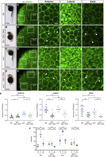
Dkk1 Sequesters β-Catenin at the Plasma Membrane (A–D) Sibling and axin1 mutant embryos at 24 h post-fertilization (hpf) (left column) and 80% epiboly (EB) with (B) and (D) or without (A) and (C) Dkk1 misexpression. 80% EB embryos were stained with an antibody against β-catenin (green) to assess endogenous β-catenin distribution in different subcellular compartments in various regions of the embryo. White-bordered squares in the second column indicate the location of single-plane confocal images in three regions of the embryo shown at a higher magnification in the three last columns. Scale bar, 20 μm. (A) Sibling embryos show patterned distribution of β-catenin (green) in different regions of the embryo at tissue and subcellular levels. In the anterior region β-catenin is detected exclusively at the plasma membrane, while high levels are seen in both nuclei and the plasma membrane, laterally. Axial cells show a low level of β-catenin in both nuclei and plasma membrane. (B) Dkk1-expressing sibling embryos display large heads and extension defects at 24 hpf and show a loss of nuclear β-catenin (green) in anterior, lateral, and axial regions of the embryo but maintain β-catenin at the plasma membrane despite the loss of cell-cell adhesion. Arrowheads point to diffuse membrane morphology. (C) axin1 null mutants show a loss of anterior fate at 24 hpf and β-catenin-positive (green) nuclei in the anterior region at 80% EB. Contrary to the current model, these gastrula embryos display the same relative subcellular distribution of β-catenin found in normal siblings across the three areas measured. (D) Despite the expected lack of telencephalon and eyes, the Dkk1-expressing axin1 mutants show an overall AP organization similar to Dkk1-expressing embryos at 24 hpf. Similar to Dkk1-RNA-injected siblings, β-catenin was retained at the plasma membrane, and cells displayed a loss of cell-cell adhesion. Arrowheads point to diffuse membrane morphology. (E) Nucleus to plasma membrane (PM) β-catenin (green) fluorescence intensity ratios quantified in anterior, lateral, and axial cells in sibling (Sib) and axin1 mutant (axin−/−) embryos, with or without Dkk1 expression. A significantly higher proportion of β-catenin is present at the PM in Dkk1-expressing Sibs and mutants in the lateral and axial regions than control embryos. Fluorescence intensity ratios were measured in 10 cells per region in each embryo (Sib, n = 2; Sib + Dkk1 RNA, n = 2; axin−/−, n = 1; and axin−/− + Dkk1 RNA, n = 1). p > 0.05 ns,∗p < 0.05, and ∗∗∗p < 0.001. (F) Absolute levels of β-catenin nuclear expression, calculated by fluorescence intensity in 10 cells in each of the three regions per embryo. A, anterior; L, lateral; and Ax, axial. p > 0.05 ns, ∗∗p < 0.01, ∗∗∗p < 0.001.
|