FIGURE

Figure 3.

ID
ZDB-FIG-191230-1203
Publication
Ge et al., 2019 - Strong static magnetic field delayed the early development of zebrafish
Other Figures
All Figure Page
Back to All Figure Page
Figure 3.

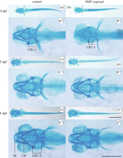
Alcian blue staining of the seven pharyngeal arches of zebrafish embryos from 4 to 6 dpf. (a,a′) Control embryo formed five ceratobranchial (CB) bones at 4 dpf. (b,b′) SMF-exposed embryo developed only three CB bones. (c,c′,d,d′) Pharyngeal arches of 5 dpf control (c,c′) and SMF-exposed (d,d′) embryos displayed no significant difference. (e,e′,f,f′) Pharyngeal arches of 6 dpf control (e,e′) and SMF-exposed (f,f′) embryos displayed no significant difference. The seven pharyngeal arches are Meckel's cartilage (M), ceratohyal (CH) bone and the five pairs of ceratobranchial (CB1–5) bones as illustrated in e′. (af) use the same scale bar, 1 mm. (a′–f′) are the magnifications, respectively, and use the same scale bar, 0.5 mm.

Expression Data

Expression Detail
Antibody Labeling
Phenotype Data

Phenotype Detail
Acknowledgments
This image is the copyrighted work of the attributed author or publisher, and ZFIN has permission only to display this image to its users. Additional permissions should be obtained from the applicable author or publisher of the image. Full text @ Open Biol.