IMAGE

Figure 3.

ID
ZDB-IMAGE-191230-1214
Source
Figures for Ge et al., 2019
Image
Figure Caption

Figure 3.

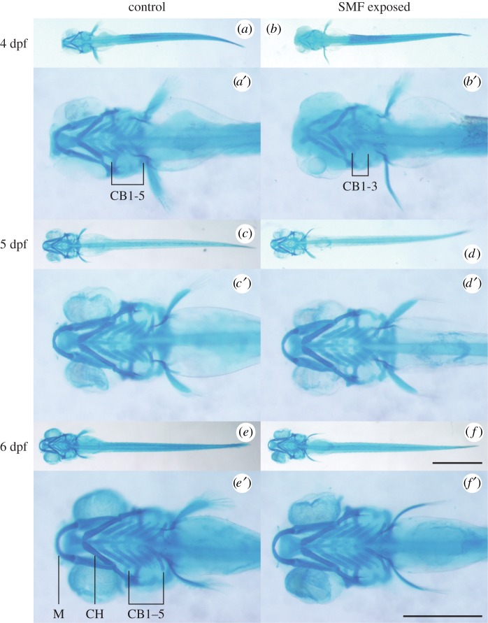
Alcian blue staining of the seven pharyngeal arches of zebrafish embryos from 4 to 6 dpf. (a,a′) Control embryo formed five ceratobranchial (CB) bones at 4 dpf. (b,b′) SMF-exposed embryo developed only three CB bones. (c,c′,d,d′) Pharyngeal arches of 5 dpf control (c,c′) and SMF-exposed (d,d′) embryos displayed no significant difference. (e,e′,f,f′) Pharyngeal arches of 6 dpf control (e,e′) and SMF-exposed (f,f′) embryos displayed no significant difference. The seven pharyngeal arches are Meckel's cartilage (M), ceratohyal (CH) bone and the five pairs of ceratobranchial (CB1–5) bones as illustrated in e′. (af) use the same scale bar, 1 mm. (a′–f′) are the magnifications, respectively, and use the same scale bar, 0.5 mm.

Acknowledgments
This image is the copyrighted work of the attributed author or publisher, and ZFIN has permission only to display this image to its users. Additional permissions should be obtained from the applicable author or publisher of the image. Full text @ Open Biol.