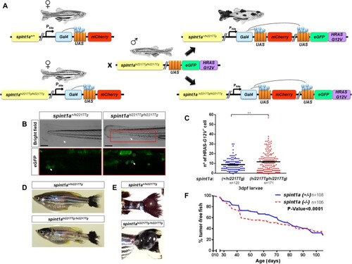
Inflammation accelerates the onset of SKCM in zebrafish. a Schematic diagram of the generation of the SKCM model line in zebrafish with Spint1a deficiency. b and c Representative images (b) and number of early oncogenically transformed eGFP-HRAS-G12V+ cells in the boxed area (c) in Spint1a-deficient larvae and control siblings at 3 dpf. Note the morphological alterations observed in the inflamed skin of the mutants (white arrows). eGFP-HRAS-G12V+ goblet cells are marked with white arrows. Scale bar 250 μm. Each point on the scatter plot represents one larva and the mean ± SEM is also shown. ** p < 0.05 according to an unpaired Student t test with Welch’s correction. d-f Impact of Spint1a deficiency on SKCM onset in zebrafish. Representative images of whole fish (d) and of nodular tail tumors (e), and Kaplan-Meier curve showing the percentage of SKCM-free fish in control and Spint1a-deficient adult fish (f). p < 0.0001 according to a Log rank Mantel-Cox test; Hazard ratio = 0.7962; 95% CI of ratio = 0.6834–0.9056
|
