
Fig. 5
- ID
- ZDB-FIG-191021-11
- Publication
- Mackin et al., 2019 - Endocrine regulation of multichromatic color vision
- Other Figures
- All Figure Page
- Back to All Figure Page
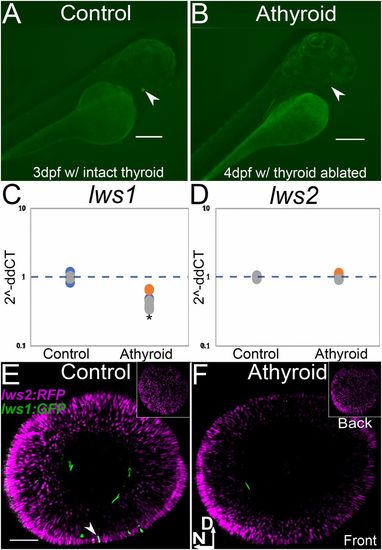
TH LOF by thyroid ablation suppresses lws1 expression at time of native onset of expression. (A and B) Transgenic [Tg(tg:nVenus-2a-nfnB)wp.rt8 that allows for thyroid ablation mediated by metronidazole treatment and nitroreductase expressing thyroglobulin cells] with intact thyroid, indicated by Venus (YFP) expression in embryo (A, arrowhead) and thyroid ablation, indicated by absence of Venus (YFP) (arrowhead shows normal location of thyroid gland) after 24-h treatment with metronidazole (B). (Scale bars in A and B, 250 μm.) (C and D) qPCR abundance of lws1 (C) and lws2 (D) transcripts (fold-change, 2−ddCT) in 6 dpf DMSO (control) and athyroid larvae. In comparison to control (n = 3) samples, lws1 is reduced in athyroid larvae (n = 3) P = 0.04 (C), and lws2 is not changed in athyroid larvae (n = 3) P = 0.32 (D). P values were calculated by comparing the ddCT values for the thyroid-ablated groups vs. controls from each experiment using a Wilcoxon Mann–Whitney U test. Statistical notation: *P < 0.05. (E and F) lws:PAC(H) lws reporter transgenic, whole-mounted eyes visualized by confocal microscopy of 6 dpf DMSO (control) (E) and athyroid larvae (F). Insets show views of the back of the eye; arrowhead in E indicates an lws1 (GFP+)-expressing cone; D, dorsal; N, nasal. (Scale bar in E [applies to E and F], 50 μm.) |