
Fig. 3
- ID
- ZDB-FIG-190918-9
- Publication
- Hendrick et al., 2019 - Bar, stripe and spot development in sand-dwelling cichlids from Lake Malawi
- Other Figures
- All Figure Page
- Back to All Figure Page
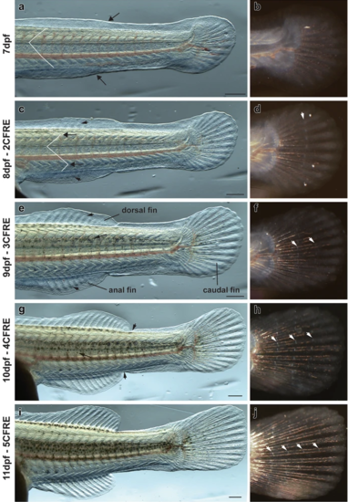
Morphological changes in median fins and myotomes during metamorphosis in D. compressiceps. aBulges (arrows) in embryonic median fin prefigure the posterior ends of the dorsal and anal fin, respectively. “V” shape of myotomes indicated in white. b Unsegmented caudal fin ray elements. cDorsal- and anal-fin ray condensations indicated by short arrows. Chevron shape of myotomes indicated by long arrows. d 2 caudal fin ray elements separated by one joint (white arrow). e Fin ray elements lengthen in dorsal fin and anal fin (short arrows). Melanophores appear on dorsal neural tube (long arrow). f 3 caudal fin ray elements separated by two joints (white arrows). g Dorsal and anal fins separated from caudal fin (short arrows). Melanophores appear in flank skin. h 4 caudal fin ray elements separated by three joints (white arrows). i Progressive disappearance of larval fin tissue from dorsal and ventral caudal fin peduncle. j 5 caudal fin ray elements separated by four joints (white arrows). The timing of metamorphosis is the same in C. azureus as in D. compressiceps. Scale = 500 µm |