Fig. 5
- ID
- ZDB-FIG-190820-22
- Publication
- Adler et al., 2019 - Clostridium perfringens Epsilon Toxin Compromises the Blood-Brain Barrier in a Humanized Zebrafish Model
- Other Figures
- All Figure Page
- Back to All Figure Page
|
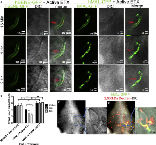
Active ETX Leads to Vessel Stenosis and Perivascular Edema (A) Zoomed-in confocal images of cerebral central arteries (CCtAs) in hBENE-GFP- and hMAL-GFP-expressing fish 15 min, 1 h, and 2 h post injection with active toxin. Green channels depict receptor expression in these vessels, gray channel depicts differential interference contrast images, “merge” overlays both images. Scale bar, 10 μm. (B) Quantitative comparison of CCtA vessel diameter over time for each condition; hMAL CCtA exposed to active ETX are significantly narrower than hBENE CCtAs exposed to ETX or hMAL CCtAs exposed to H149A mutant pETX starting at 1 HPI and showed significant decrease from 15 MPI to 2 HPI (two-way ANOVA with Tukey's test, *p < 0.05, **p < 0.01). Data are represented as mean ± SEM. (C) Confocal differential interference contrast macroscopic z-slice of zebrafish brain exhibiting perivascular edema in an hMAL-GFP-expressing fish and (C′) zoomed in single vessel. (D) Image (C) with merged hMAL-GFP and dextran channels and (D′) merged, zoomed-in single vessel. Scale bar, 25 μm for full image and 10 μm for zoomed-in vessel. |

