
FIGURE
Fig. 7
- ID
- ZDB-FIG-190816-9
- Publication
- Kuroda et al., 2018 - Roles of basal keratinocytes in actinotrichia formation
- Other Figures
- All Figure Page
- Back to All Figure Page
Fig. 7
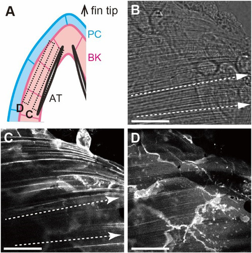
In vivo orientation of the basal keratinocyte membrane in the larval fin. (A) Schematic of the horizontal section of larval fin. The top shows the fin tip and the two boxed regions correspond to the cross section of (C) and (D). AT, BK, and PC correspond actinotrichia, basal keratinocyte and peridermal cell respectively. (B) Phase contrast image of the fin tip in. |
Expression Data
Expression Detail
Antibody Labeling
Phenotype Data
Phenotype Detail
Acknowledgments
This image is the copyrighted work of the attributed author or publisher, and
ZFIN has permission only to display this image to its users.
Additional permissions should be obtained from the applicable author or publisher of the image.
Reprinted from Mechanisms of Development, 153, Kuroda, J., Iwane, A.H., Kondo, S., Roles of basal keratinocytes in actinotrichia formation, 54-63, Copyright (2018) with permission from Elsevier. Full text @ Mech. Dev.