
Fig. 3
- ID
- ZDB-FIG-190808-5
- Publication
- Cosacak et al., 2019 - Single-Cell Transcriptomics Analyses of Neural Stem Cell Heterogeneity and Contextual Plasticity in a Zebrafish Brain Model of Amyloid Toxicity
- Other Figures
- All Figure Page
- Back to All Figure Page
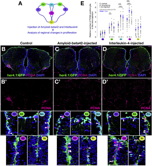
Aβ42 and IL-4 Affect Specific Progenitor Populations in the Adult Zebrafish Brain (A) Schematic view of regionalization in the adult zebrafish telencephalon. (B–D’) Immunohistochemistry (IHC) staining for her4.1-driven GFP and PCNA on control (B), amyloid-injected (C), and IL-4-injected (D) telencephalic sections. In (B’), (C’), and (D’), PCNA channels are shown. Close-up views of GFP and PCNA double IHC are shown below the single channels. Regions are marked with color-codes as in (A). Scale bars, 50 μm. (E) Quantification graph for the relative number of proliferating progenitors. Three brains were used for every experimental group. Data are represented as mean ± SEM. The levels of significance are ∗p < 0.05, ∗∗p < 0.01, and ∗∗∗p < 0.001. |