
FIGURE
Fig. 4
- ID
- ZDB-FIG-190807-28
- Publication
- Mishra et al., 2019 - Zebrafish model of amyloid light chain cardiotoxicity: regeneration vs degeneration
- Other Figures
- All Figure Page
- Back to All Figure Page
Fig. 4
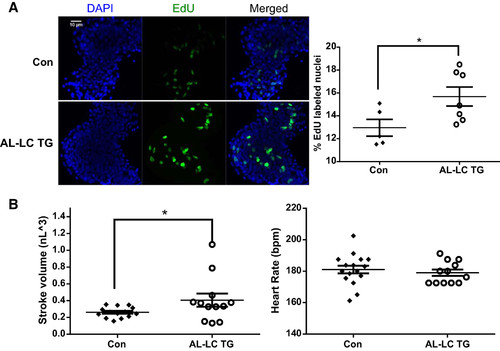
Early signs of functional as well as cellular compensation begin in AL-LC transgenic fish embryos. Increased proliferation as indicated by increased EdU-labeled nuclei (green) were detectable in the 48 hpf transgenic zebrafish hearts compared with control hearts (*P = 0.04) (A). Dimensional analysis to measure cardiac function was performed at 48 hpf as previously described in (23) revealing an increase in stroke volume (*P = 0.03, left) while maintaining a comparable heart rate (B). bpm, beats/min; Con, control; EdU, 5-ethynyl-2′-deoxyuridine; hpf, hours postfertilization; TG, transgenic.
|
Expression Data
Expression Detail
Antibody Labeling
Phenotype Data
| Fish: | |
|---|---|
| Observed In: | |
| Stage: | Long-pec |
Phenotype Detail
Acknowledgments
This image is the copyrighted work of the attributed author or publisher, and
ZFIN has permission only to display this image to its users.
Additional permissions should be obtained from the applicable author or publisher of the image.
Full text @ Am. J. Physiol. Heart Circ. Physiol.