
Fig. 6
- ID
- ZDB-FIG-190806-22
- Publication
- Baumgartner et al., 2019 - Identification of regulatory elements recapitulating early expression of L-plastin in the zebrafish enveloping layer and embryonic periderm
- Other Figures
- All Figure Page
- Back to All Figure Page
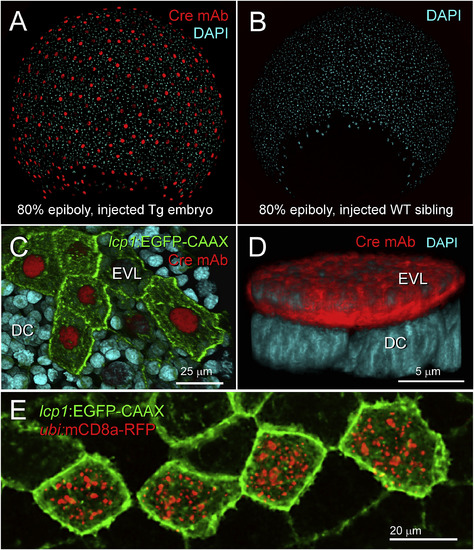
Expression and activity of Cre recombinase in lcp1(Cre-P2A-EGFP-CAAX) transgenic embryos. A. Fixed transgenic embryo at 80% epiboly. Immunohistochemical labeling shows robust nuclear signal in all EVL cells. red = anti-Cre monoclonal antibody, blue = DAPI nuclear stain. B. Fixed wild type sibling embryo co-stained with the specimen in (A). Although exposed to the same primary and secondary antibody solutions, no nuclei stain red. C. Fixed transgenic embryo, animal cap at 80% epiboly. EGFP and Cre signals are positively correlated. Cells with bright membranes have bright nuclei; cells with dim membranes have dim nuclei. D. High magnification view of a single, Cre-positive EVL nucleus (red) overlying multiple Cre-negative deep cell nuclei (blue). E. In vivo confirmation of Cre activity. After transient transgenesis with a Cre reporter plasmid, a live cluster of transgenically labeled periderm cells (green) expresses recombinant RFP-tagged mouse CD8a (red). For details, see Methods. |
Reprinted from Gene expression patterns : GEP, 32, Baumgartner, E.A., Compton, Z.J., Evans, S., Topczewski, J., LeClair, E.E., Identification of regulatory elements recapitulating early expression of L-plastin in the zebrafish enveloping layer and embryonic periderm, 53-66, Copyright (2019) with permission from Elsevier. Full text @ Gene Expr. Patterns