
Fig. 3
- ID
- ZDB-FIG-190806-19
- Publication
- Baumgartner et al., 2019 - Identification of regulatory elements recapitulating early expression of L-plastin in the zebrafish enveloping layer and embryonic periderm
- Other Figures
- All Figure Page
- Back to All Figure Page
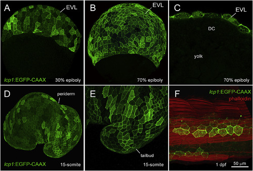
Transgenic expression in lcp1:EGFP-CAAX embryos. A. 30% epiboly, transgenic embryo in equatorial view. The EVL shows weak, variegated EGFP expression. B. 70% epiboly, transgenic embryo in equatorial view. Signal intensifies as the EVL spreads over the yolk. C. 70% epiboly, cutaway view of a single focal plane. Only EVL cells are EGFP+, in contrast to the deep cells (DC) and yolk syncytial layer (YSL). D. Live embryo at the 15-somite stage. lcp1:EGFP-CAAX labels the periderm, the outermost epithelial layer. E. Magnification of the tailbud in D, showing variegated EGFP in adjacent peridermal cells. F. Fixed embryo at 1 dpf, with phalloidin-568 counterstain. A patch of label-retaining peridermal cells (green) lies over the developing trunk musculature (red). |
Reprinted from Gene expression patterns : GEP, 32, Baumgartner, E.A., Compton, Z.J., Evans, S., Topczewski, J., LeClair, E.E., Identification of regulatory elements recapitulating early expression of L-plastin in the zebrafish enveloping layer and embryonic periderm, 53-66, Copyright (2019) with permission from Elsevier. Full text @ Gene Expr. Patterns