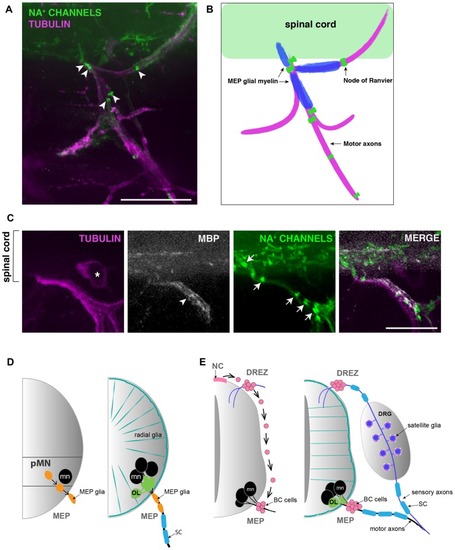
MEP glial myelin is flanked by nodes of ranvier. (A) Lateral view of a 5 dpf zebrafish trunk stained with antibodies specific to pan Na+ channels (clone k58/35) and acetylated tubulin shows that MEP glial territory is flanked by sodium channels clustered in Nodes of Ranvier (arrowheads) along motor nerve roots. (B) Diagram showing MBP+ MEP glial myelin (blue) delineated by nodes of Ranvier (green) along motor nerve root axons (magenta). (C) Immunostaining showing MBP+ MEP glial myelin (arrowhead) and nodes of Ranvier (arrows) along motor nerve root axons (magenta). Asterisks point to dorsal root ganglion (DRG). (D) Schematic of zebrafish MEP transition zones (TZs) and the diverse populations of glial cells orchestrating the CNS/PNS boundary. CNS-derived MEP glia (orange) that reside along motor neuron (mn; black) axons restrict the oligodendrocyte lineage (OL; green) to the spinal cord. Radial glia (teal green) cover the surface of the spinal cord and prevent peripheral glia such as MEP glia and SC (blue) from entering the CNS. (E) Schematic of a mammalian neural tube showing neural crest (NC)-derived boundary cap (BC) cells (pink) sitting at the dorsal root entry zone (DREZ) and MEP TZs. BC cells prevent mn (black) cell bodies from transgressing the spinal cord boundary. Scale bar, (A) 50 μm, (C) 25 μm.
|

