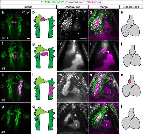
The tbx1+ cone forms the ventricular myocardium but not the BA. a, f, k, p Maximum intensity projections and schematics of representative photoconverted (a, f, k) or control (p) tbx1:Dendra2 embryos; dorsal views, anterior to the top. At 22 hpf, tbx1:Dendra2-expressing embryos were illuminated with a 405-nm laser in a confined region of interest to convert Dendra2-green to Dendra2-red in specific tbx1 reporter-expressing domains. c, h, m, r SPIM-imaged hearts and graphical representations of embryos photoconverted as in a, f, k and p and stopped from contracting with BDM at 3.5 dpf; maximum intensity projections (c, d, m, n, r, s) or optical Z-section (h, i), ventral views, anterior to the top. a–e Dendra2-red-positive sheath cells give rise to ventricular cardiomyocytes, including the SHF-derived distal ventricle, but not to the BA. The red signal within the BA (b) derives from autofluorescent blood also detected in non-photoconverted tbx1:Dendra2 embryos (see p–t). f–j Medial migrating cells posterior to the cardiac cone contribute to the most distal myocardium at the dorsal side of the heart (arrowhead in h, i) and to a proximal portion of the BA in 3/3 analyzed embryos. k–o Photoconversion of a broad area in the tbx1:Dendra2-positive pharyngeal ALPM posterior to and on the right of the linear heart tube marks the right side of the BA (dotted outline in n). p–t Red signals in the chambers and on top of the pericardium are due to unspecific autofluorescence also detected in controls. Scale bars 50 µm
|
