
Figure 5
- ID
- ZDB-FIG-190723-1488
- Publication
- Zhu et al., 2019 - Mutant Ahi1 Affects Retinal Axon Projection in Zebrafish via Toxic Gain of Function
- Other Figures
- All Figure Page
- Back to All Figure Page
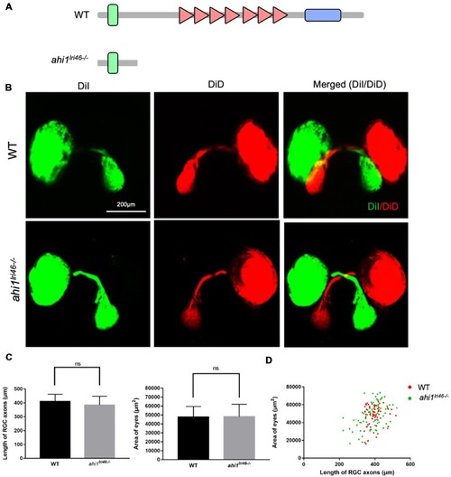
The ahi1 knockout (KO) line ahi1lri46−/−embryos exhibited no significant difference from WT in optic nerve projection length and eyes size. (A) The ahi1lri46−/− zebrafish has depleted most of the region of the ahi1 gene and only retains the N-terminal coiled-coil domain. (B) Fluorescent images of WT and ahi1lri46−/−zebrafish optic nerve projections at 4 dpf. Scale bar: 200 μm. (C) The quantified optic nerve projection length and eye sizes of WT and ahi1lri46−/− embryos. (D) The two-dimensional scatter diagram for the eye size of WT and ahi1lri46−/− embryos; the X-axis represents the length of optic nerve projection (μm2), and the Y-axis represents the size of eyes (μm2). ns, not significant. Error bars denote SEM. |
| Fish: | |
|---|---|
| Observed In: | |
| Stage: | Day 4 |