
Figure 3
- ID
- ZDB-FIG-190723-1154
- Publication
- Rausch et al., 2018 - The Hippo Pathway Regulates Caveolae Expression and Mediates Flow Response via Caveolae
- Other Figures
- All Figure Page
- Back to All Figure Page
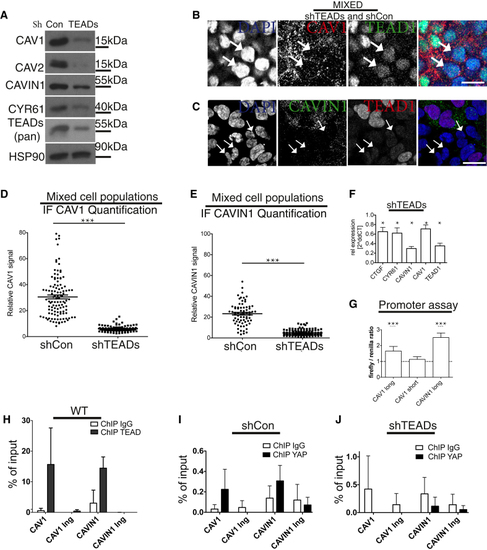
(A) Western blots of lysates from shTEADs and shCon HEK293A cells. (B) Mixed cell population of shTEADs and shCon HEK293A cells labeled for DAPI (blue), CAV1 (red), and TEAD1 (green). Discrete cell populations are shown in (C) Mixed cell population of shTEADs and shCon HEK293A cells labeled for DAPI (blue), TEAD1 (red), and CAVIN1 (green). Discrete cell populations are shown in Arrows in (B) and (C): examples of shTEADs cells. Scale bars in (B) and (C) represent 15 μm. (D and E) Dot plot of CAV1 levels from images, as shown in (B) ( (F) qPCR data from cells as in (A) (related to (G) YAP drives (H) Real-time PCR analysis of TEAD1 chromatin immunoprecipitation (ChIP) in HEK293A cells. The precipitated DNA was quantitated using primers specific for a promoter region or a control in-gene (Ing) region. Data are means ± SD of triplicates from a representative experiment. Endogenous TEAD1 binds to both (I and J) YAP ChIP for |