|
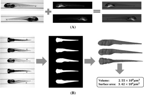
Typical applications of zebrafish segmentation. a Fluorescence images visualisation and evaluation. Bright-field zebrafish images offer reference for the shape of the specimen (column one). Fluorescent images present informative signals, e.g. the blood vessels in green (column two). Accurate segmentation of the bright-field image provides a good shape reference to evaluate the fluorescent signals, for example, the development and concentration of specific cells (column three). b 3D zebrafish reconstruction from axial views. Axial-view zebrafish images (column one) are segmented to obtain 2D binary shapes (column two), from which the axial-view-based 3D reconstruction produces 3D models as well as 3D measurements (column three) (colour figure online)
|