
Fig. 6
- ID
- ZDB-FIG-190701-29
- Publication
- Posner et al., 2019 - Why does the zebrafish cloche mutant develop lens cataract?
- Other Figures
- All Figure Page
- Back to All Figure Page
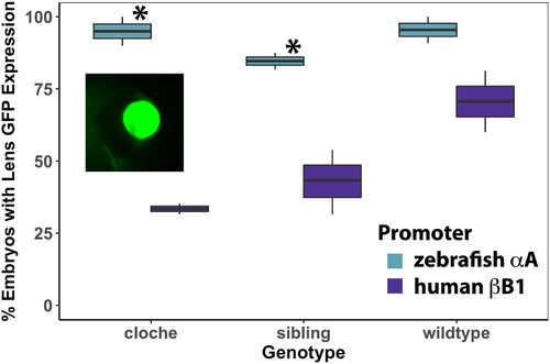
Percent of embryos with lens GFP expression after injection of zebrafish αA-crystallin promoter/GFP and human βB1-crystallin promoter/GFP plasmids. Data show that the native αA-crystallin promoter drives greater GFP expression in lens compared to the human bB1 promoter in cloche and non-phenotype siblings (Yates Corrected X2 test: X2 = 16.85, p value<0.001; X2 = 21.38, p value<0.001 respectively), but this difference was not statistically significant in wildtype embryos (X2 = 3.35, pvalue>0.05). Each box and whisker blot represents two independent experiments (except for the βB1 sibling value which included three independent experiments). Each independent experiment included between 3 and 54 embryos at 4 dpf (median = 19). Inset shows an example of GFP lens expression in a cloche embryo produced by the αA-crystallin promoter. |