
Fig. 3-S2
- ID
- ZDB-FIG-190628-24
- Publication
- Giovannone et al., 2019 - Programmed conversion of hypertrophic chondrocytes into osteoblasts and marrow adipocytes within zebrafish bones
- Other Figures
- All Figure Page
- Back to All Figure Page
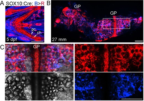
Neural crest contributions to the Ch bone and marrow adipocytes.(A) In SOX10:Cre; bactin2:loxP-tagBFP-stop-loxP-DsRed animals, the human SOX10 promoter drives Cre expression and subsequent recombination of tagBFP (blue) to DsRed (red) in neural crest lineage cells (as opposed to the zebrafish sox10 promoter of Figure 3, which has additional later expression throughout chondrocytes of both crest and mesoderm origin). Confocal projection of a zebrafish embryo (five dpf) shows nearly all of the chondrocytes (ch) and perichondral cells (pc) are labeled red. n = 4. (B) Confocal projection of a dissected Ch bone from an adult zebrafish (27 mm SL). Nearly all growth plate (GP) chondrocytes are labeled red and numerous red lineage cells are seen throughout the marrow cavity and along the cortical surface. n = 4. (C) Merged and separate channels corresponding to the boxed region in (B), which is centered on the growth plate. The brightfield channel shows the many lipid droplets characteristic of adipocytes. The red channel shows the circular profile of the cytoplasm surrounding the lipid droplets in labeled adipocytes, as well as numerous smaller mesenchymal cells, some of which are likely osteoblasts on the cortical surface. The blue channel shows the presence of cells of non-neural-crest-origin, such as endothelial cells of mesoderm origin that form the vasculature within Ch (see Figure 2). Similar contributions were observed in four independent animals. Scale bars = 50 μm (A), 200 μm (B,C). |