
Fig. 4
- ID
- ZDB-FIG-190627-15
- Publication
- Jung et al., 2019 - Znf76 is associated with development of the eyes, midbrain, MHB, and hindbrain in zebrafish embryos
- Other Figures
- All Figure Page
- Back to All Figure Page
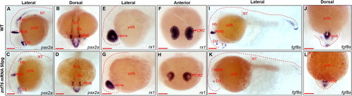
Marker analysis in znf76-overexpressing embryos WISH was performed at 24 hpf in znf76-overexpressing embryos with pax2a, rx1, and fgf8a to investigate the morphological defects at a molecular level. Lateral view (A, C, E, G, I, and K); anterior view (B, D, F, H, J, and L). pax2a transcripts were significantly reduced in the optic stalk, MHB, and hindbrain neurons compared with the WT (A-D). rx1 marks neural cells in the retina of WT embryos at 24 hpf (E,F). rx1transcripts were reduced in znf76-overexpressing embryos in the retina, especially in the CMZ area of the eyes (F,H); the size of the eyes was also reduced compared with the WT (E,G). fgf8a is a marker for the dorsal diencephalon, MHB, posterior somites, and posterior notochord. At 24 hpf, there was a marked reduction in fgf8a transcripts in these regions in znf76-overexpressing embryos (K,L) compared with the WT (I,J). CMZ – central marginal zone, Dd – dorsal diencephalon, MHB – midbrain–hindbrain boundary, Hb – hindbrain, T – telencephalon, NT – neural tube, OS – optic stalk. Scale bar: 100 µm. |