
Fig. 5
- ID
- ZDB-FIG-190606-8
- Publication
- Kelu et al., 2018 - Characterization of ADP-ribosyl cyclase 1-like (ARC1-like) activity and NAADP signaling during slow muscle cell development in zebrafish embryos
- Other Figures
- All Figure Page
- Back to All Figure Page
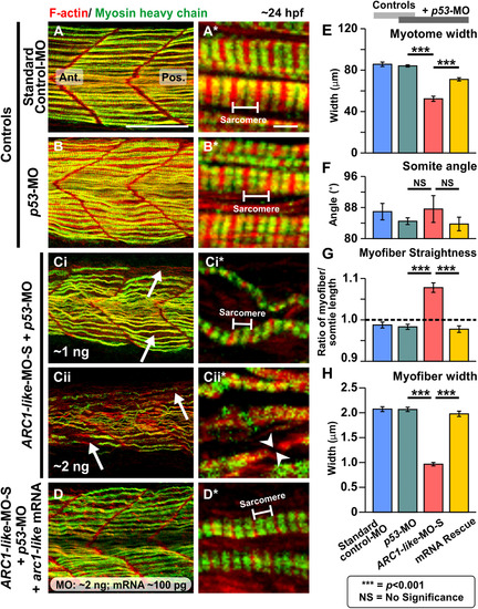
Effect of MO-based knockdown of ARC1-like (without and with mRNA rescue) on the organization of the trunk musculature and the formation of the sarcomeres. Embryos were injected with (A) ~ 5 ng standard control-MO; (B) ~ 5 ng p53-MO; (Ci) ~ 1 ng ARC1-like-MO-S + ~ 1 ng p53-MO; (Cii) ~ 2 ng ARC1-like-MO-S + ~ 2 ng p53-MO; or (D) ~ 2 ng ARC1-like-MO-S + ~ 2 ng p53-MO + ~ 100 ng arc1-like mRNA. All the embryos were fixed at ~ 24 hpf and dual-labelled with phalloidin and the F59 antibody, to visualize F-actin (in red) and myosin heavy chain (in green) in the trunk musculature, respectively. The panels show a series of optical sections projected as single images at (A, B, Ci, Cii, D) low and (A*, B*, Ci*, Cii*, D*)higher magnification when the red and green channels are merged; overlapping regions are shown in yellow. The higher magnification images of the slow myofibers reveal the presence or absence of the sarcomeric banding pattern of the F-actin and myosin heavy chain. The white arrows in panels (Ci, Cii) show the disorganization of some of the slow myofibers, whereas the white arrowheads in panel (Cii*) indicate a gap in one of the myofibers. Ant. and Pos. are anterior and posterior, respectively. Scale bars, 50 µm (in panels A, B, Ci, Cii, D), and 2 µm (in panels A*, B*, Ci*, Cii*, D*). (E–H) In order to determine the level of disruption on slow muscle cell development following ARC1-like-knockdown, various dimensions of the trunk musculature and slow myofibers were measured. These bar graphs show the mean ± SEM: (E) myotome width (all n = 18, from 6 embryos), (F)somite angle (n = 18, from 6 embryos), (G) myofiber length:somite length ratio (n = 162 from 6 embryos); and (H)myofiber width (n = 60, from 6 embryos). The black dashed line in panel (G) indicates a myofiber:somite length ratio of 1. The asterisks indicate statistically significant differences at p < 0.001 (***), whereas NS indicates that no significant difference was observed. |
| Antibody: | |
|---|---|
| Fish: | |
| Knockdown Reagents: | |
| Anatomical Term: | |
| Stage: | Prim-5 |
| Fish: | |
|---|---|
| Knockdown Reagents: | |
| Observed In: | |
| Stage: | Prim-5 |
Reprinted from Developmental Biology, 445(2), Kelu, J.J., Webb, S.E., Galione, A., Miller, A.L., Characterization of ADP-ribosyl cyclase 1-like (ARC1-like) activity and NAADP signaling during slow muscle cell development in zebrafish embryos, 211-225, Copyright (2018) with permission from Elsevier. Full text @ Dev. Biol.