Fig. 4
- ID
- ZDB-FIG-190604-64
- Publication
- Sinnberg et al., 2018 - Wnt-signaling enhances neural crest migration of melanoma cells and induces an invasive phenotype
- Other Figures
- All Figure Page
- Back to All Figure Page
|
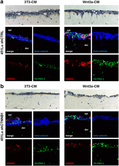
Wnt3a induces invasive growth of melanoma cells in organotypic tissue skin reconstructs (TSR). a 451 LU melanoma cells were seeded together with HaCat epidermal cells onto a layer of collagen I embedded human fibroblasts. TSR exposed to Wnt3a conditioned medium (Wnt3a-CM) showed a pronounced invasive morphology in the H&E staining (upper pictures) when compared to cells exposed to control medium (3T3-CM). Immunofluorescence stainings for HMB45 (red) and beta-catenin (blue) identified melanoma cells (HMB45+), revealed beta-catenin expression levels and verified the invasion of single 451 LU cells from the epidermis (epi) into the dermal part (der). Nuclei were stained with YO-PRO-1 (green). b Knockdown of beta-catenin (blue) with shRNA (shCTNNB1) reduced the invasion of 451 LU melanoma cells (HMB45+, red) into the dermal part of the TSR |

