
- Title
-
Wnt-signaling enhances neural crest migration of melanoma cells and induces an invasive phenotype
- Authors
- Sinnberg, T., Levesque, M.P., Krochmann, J., Cheng, P.F., Ikenberg, K., Meraz-Torres, F., Niessner, H., Garbe, C., Busch, C.
- Source
- Full text @ Mol. Cancer
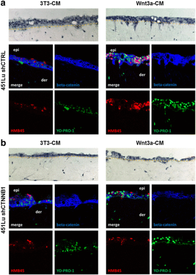
Wnt3a induces invasive growth of melanoma cells in organotypic tissue skin reconstructs (TSR). a 451 LU melanoma cells were seeded together with HaCat epidermal cells onto a layer of collagen I embedded human fibroblasts. TSR exposed to Wnt3a conditioned medium (Wnt3a-CM) showed a pronounced invasive morphology in the H&E staining (upper pictures) when compared to cells exposed to control medium (3T3-CM). Immunofluorescence stainings for HMB45 (red) and beta-catenin (blue) identified melanoma cells (HMB45+), revealed beta-catenin expression levels and verified the invasion of single 451 LU cells from the epidermis (epi) into the dermal part (der). Nuclei were stained with YO-PRO-1 (green). b Knockdown of beta-catenin (blue) with shRNA (shCTNNB1) reduced the invasion of 451 LU melanoma cells (HMB45+, red) into the dermal part of the TSR |
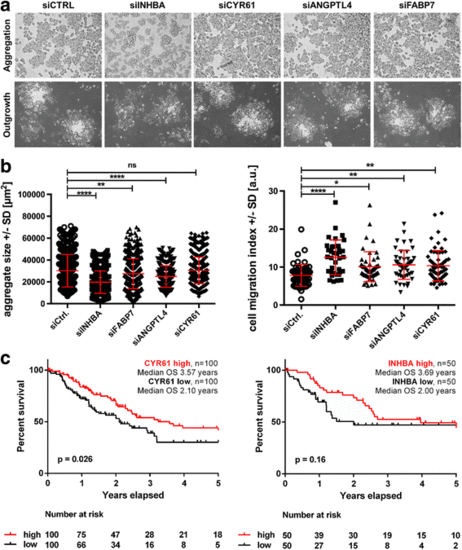
siRNA knock-down confirms the double role of genes simultaneously involved in embryonic neural crest induction and melanoma cell adhesion. a SKMEL28 cells aggregated during 24 h of roller culture after knockdown of the four gene candidates (upper row) and were microphotograped (10X magnification) Further cultivation of the aggregates for 24 h was used to detect the outgrowth of migratory cells (10X magnification). b For all four knocked-down genes a significant decrease of aggregate size (left diagram) compared to the siCtrl SKMEL28 cells was observed (ns: not significant, **: p < 0.01, ****: p < 0.0001, One way ANOVA). Depicted in red: mean aggregate size in μm2±SD. An increased cell migration index (right diagram) was detected for all four knocked-down genes in the transfected SKMEL28 cells compared to siCtrl SKMEL28 cells (*: p < 0.05, **: p < 0.01, ****: p < 0.0001, One way ANOVA). Depicted in red: mean cell migration index±SD. c Kaplan-Meier plots of CYR61- or INHBA-expression in primary melanomas demonstrating a correlation of gene expression with overall survival of melanoma-afflicted patients |