
Fig. 2
- ID
- ZDB-FIG-190530-27
- Publication
- Cao et al., 2018 - Covering and Re-Covering the Heart: Development and Regeneration of the Epicardium
- Other Figures
- All Figure Page
- Back to All Figure Page
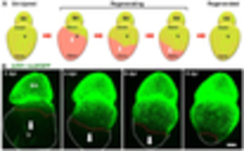
Epicardial regeneration. (A) Schematic for epicardial ablation and regeneration. The epicardium is shown in green color. The white arrows indicate the directions of epicardial cell migration; (B) explanted adult zebrafish hearts were imaged daily post epicardial ablation. The epicardial cell sheet regenerates along the ventricular surface in a base-to-apex direction (arrows). The nuclei of epicardial cells are labeled with nuclear GFP (tcf21:nucEGFP). White dashed lines outline the ventricles, and red dashed lines indicate the leading edge of the migrating epicardial cell sheet. Scale bar: 200 μm. BA, bulbous arteriosus; v, ventricle; dpi, days post injury. |