
- Title
-
Covering and Re-Covering the Heart: Development and Regeneration of the Epicardium
- Authors
- Cao, Y., Cao, J.
- Source
- Full text @ J Cardiovasc Dev Dis
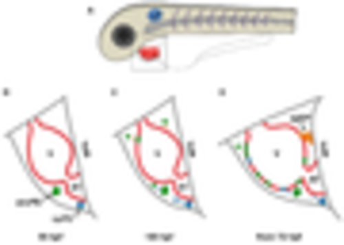
Schematic of epicardium formation in zebrafish. (A) Schematic of the anterior half of a zebrafish embryo. The framed region in (A) is enlarged to show details below (B–D); (B) at approximately 55 hpf, two PE clusters emerge from the mesothelial wall close to the atrioventricular canal (avcPE) and the venous pole PE (vpPE); (C) from approximately 65 hpf, cells are released (blue arrows) from the PE clusters and start to attach to the ventricular surface (orange arrows); (D) from 72 hpf, cells from the arterial pole epicardial precursor (apEP) pool (black arrow) are transferred to the ventricular surface through a cell bridge. V, ventricle; AT, atrium. |
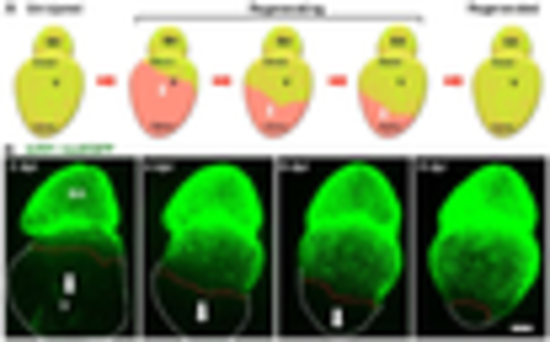
Epicardial regeneration. (A) Schematic for epicardial ablation and regeneration. The epicardium is shown in green color. The white arrows indicate the directions of epicardial cell migration; (B) explanted adult zebrafish hearts were imaged daily post epicardial ablation. The epicardial cell sheet regenerates along the ventricular surface in a base-to-apex direction (arrows). The nuclei of epicardial cells are labeled with nuclear GFP (tcf21:nucEGFP). White dashed lines outline the ventricles, and red dashed lines indicate the leading edge of the migrating epicardial cell sheet. Scale bar: 200 μm. BA, bulbous arteriosus; v, ventricle; dpi, days post injury. |